FISH Analysis on Tissue Section
- Deletion / amplification studies in tumor tissues
- Detection of chromosome translocation using break/apart probes
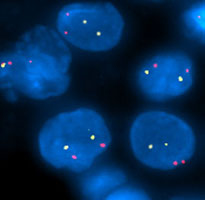
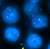
Amplification / Deletion analysis in human brain tumors using 1p32 BAC probe (yellow) and 1q44 BAC probe (red).
Normal
Loss
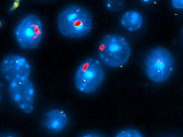
Detection of X and Y chromosomes in mouse brain tissue section
Mouse Y chromosome painting probe (red) and mouse X chromosome BAC probe (yellow) was detected in paraffin embedded mouse brain tissue section with DAPI counter stain.
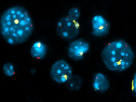
Detection of X and Y chromosomes in mouse liver tissue section
Mouse Y chromosome painting probe (yellow) and mouse X chromosome BAC probe (red) was detected in paraffin embedded mouse brain tissue section with DAPI counter stain.
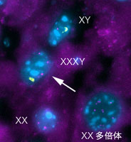
Cell fusion in Mouse liver
Y: yellow, X: purple
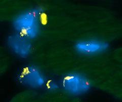
Mouse heart
Y: yellow, X: red
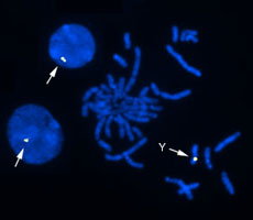
Detection of Rat Y chromosome on rat metaphase spreads using Rat Y FISH probe
Detection of Rat Y FISH probe signals on formalin fixed paraffin embedded sections of rat intestine
